Day 3 @ MBL
November 13, 2019
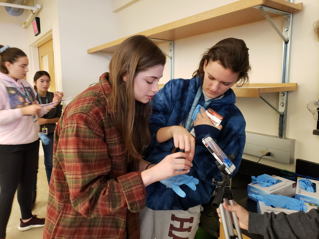
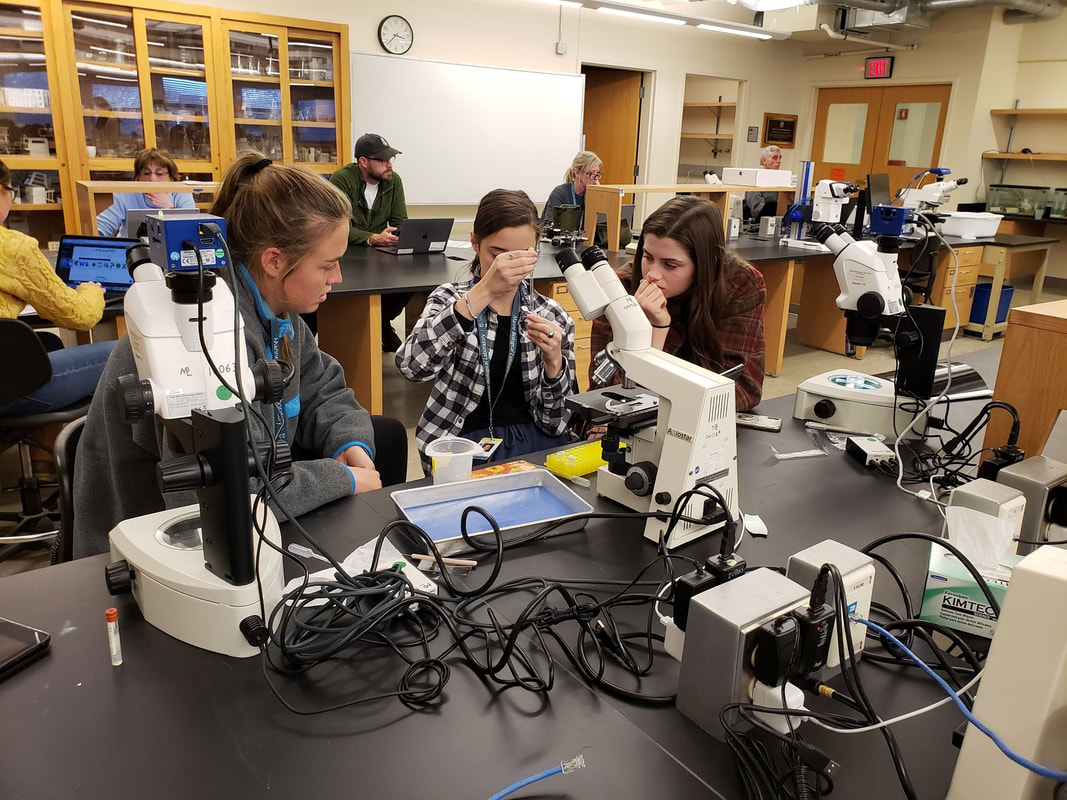
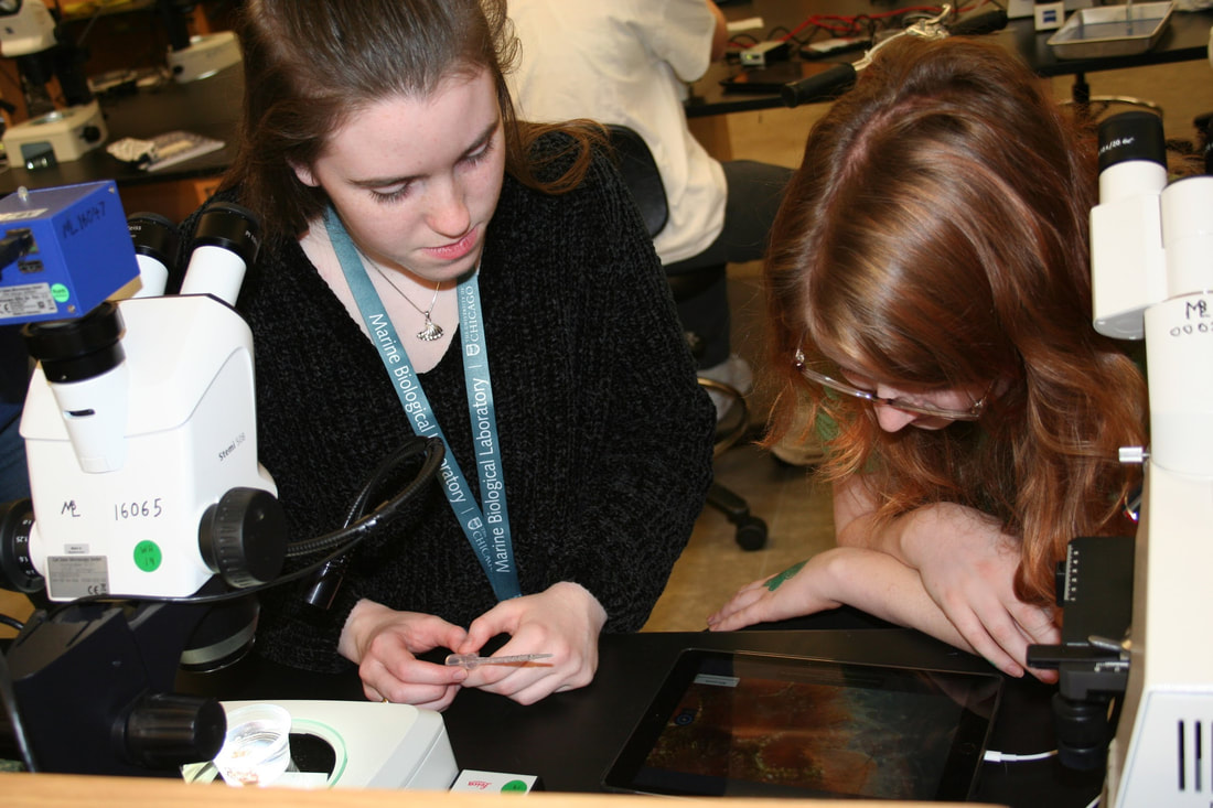
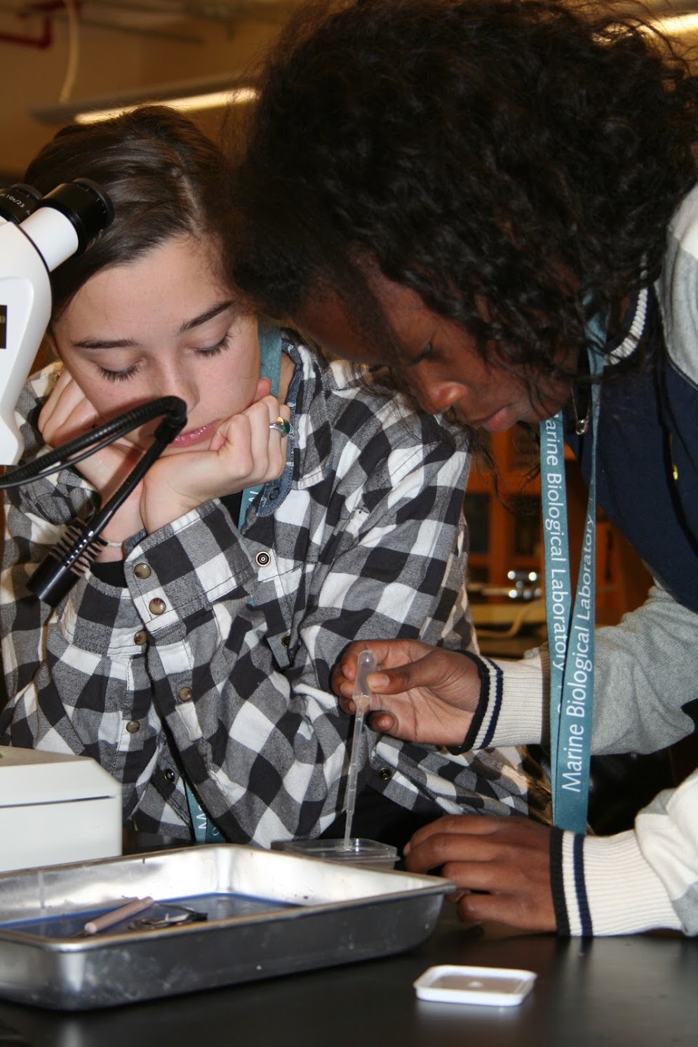
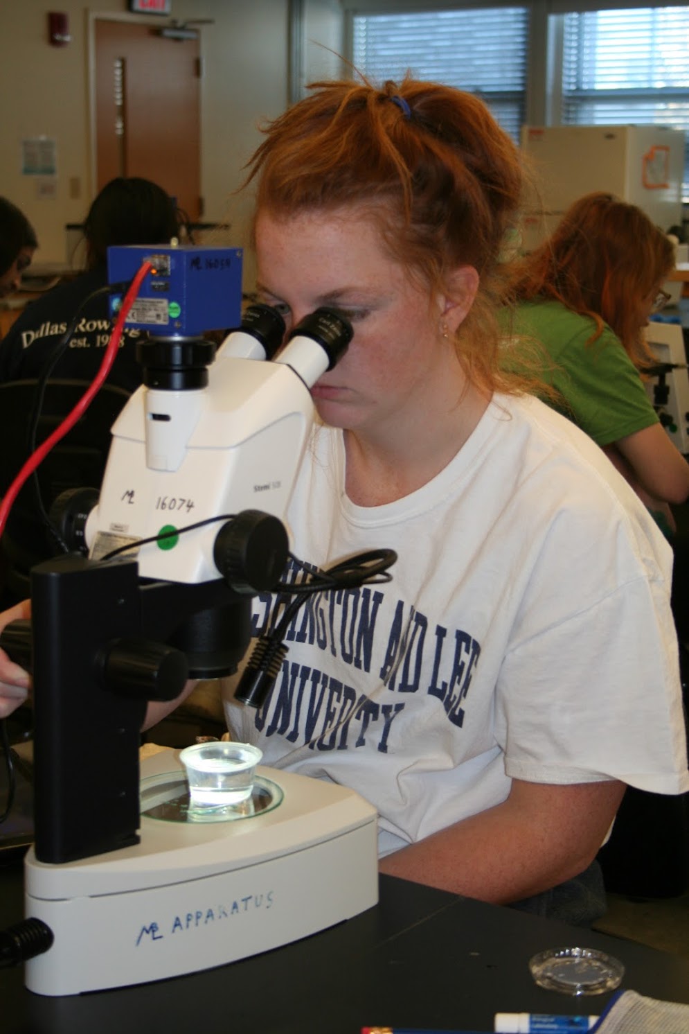
This morning was all about invertebrates and their anatomy. After an introductory lecture, we headed to the lab where we dissected a few different types of invertebrates including sea urchins, whelk, sea stars, and cephalopods. This was a great opportunity to compare and contrast different types of invertebrate anatomy and physiology. One particular difference between various species of invertebrates is that some have a closed circulatory system while others have an open one. We also had a chance to observe live horseshoe crabs. Their blood, which is blue because it contains copper, is used to detect biological endotoxins in medical applications.
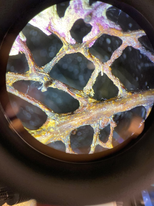
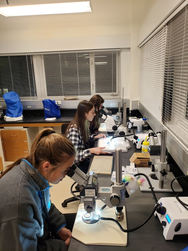
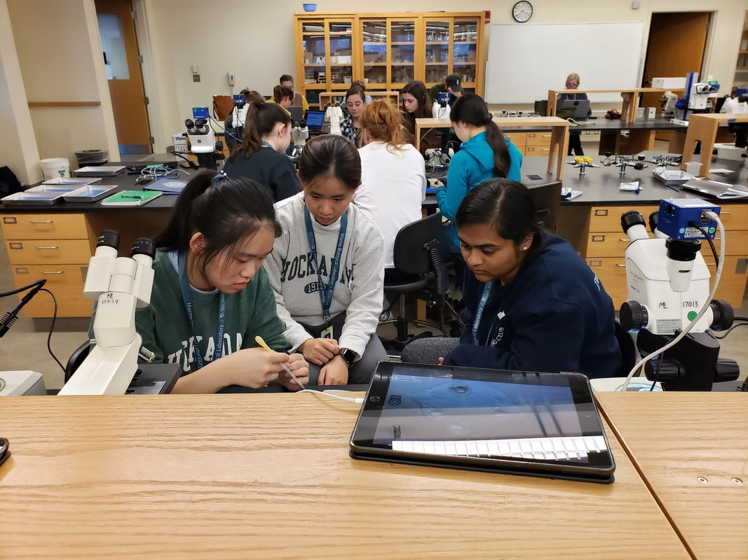
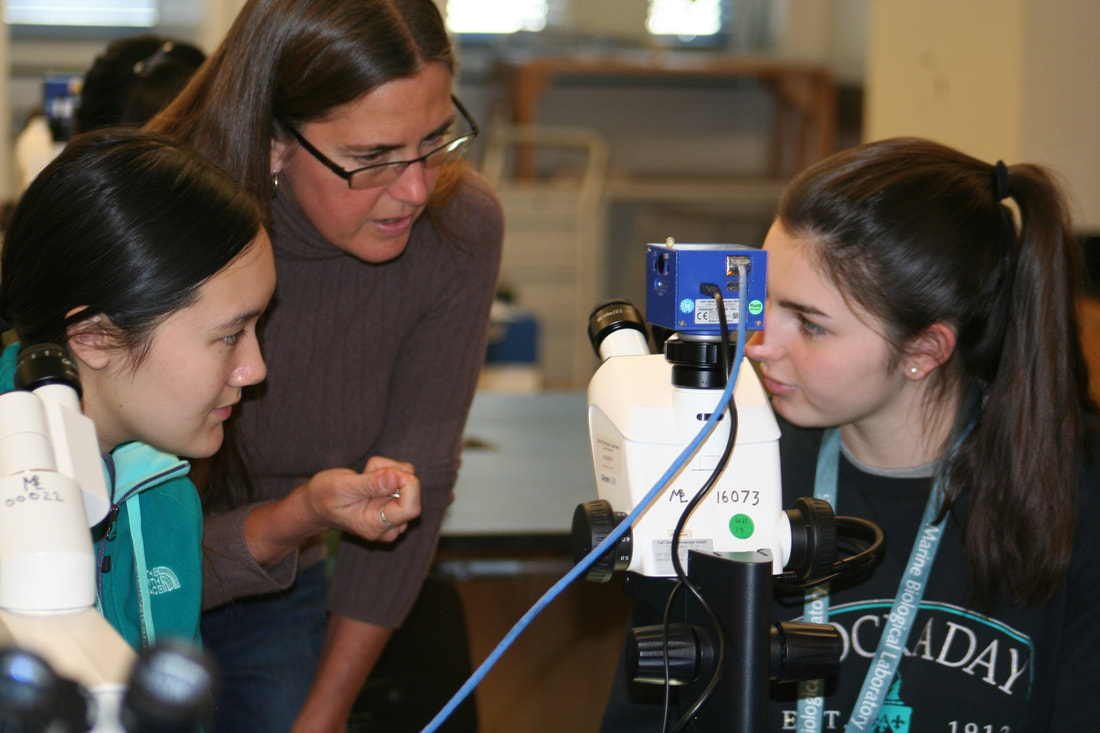
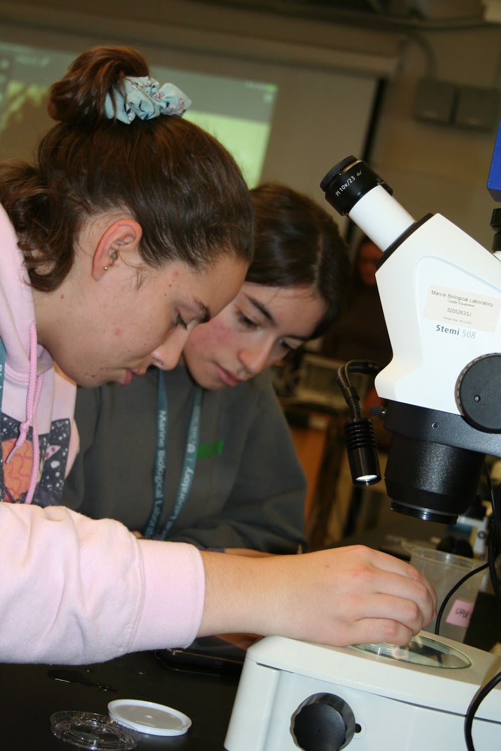
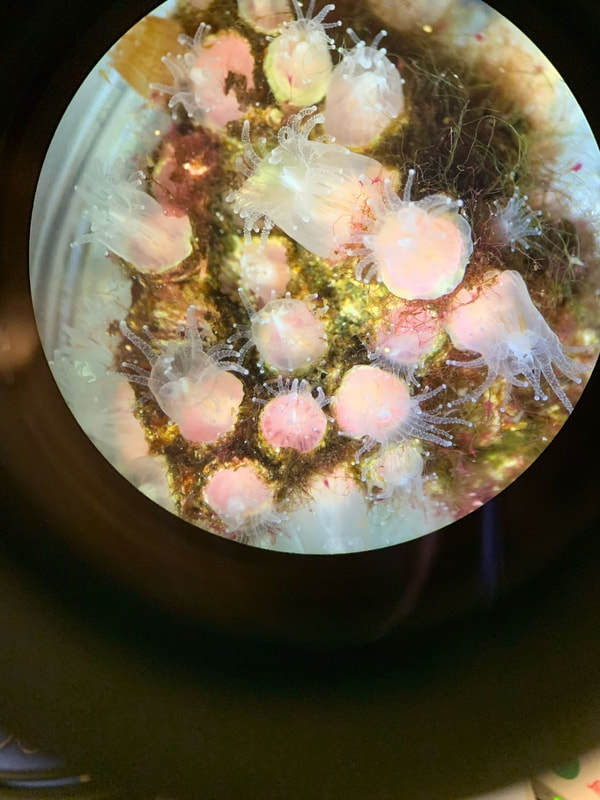
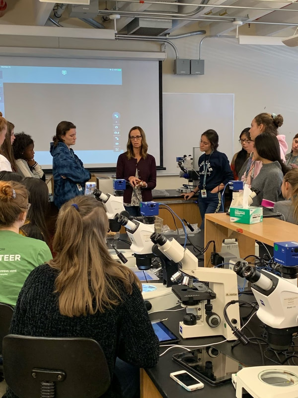
After lunch, our focus shifted to coral anatomy, physiology, reproduction, and disease. Dr. Loretta Roberson, who is a full-time researcher here at the MBL, gave a lecture on this topic and then led a lab section where we had the chance to observe different corals and anemone. The highlight of this was getting to feed brine shrimp to the sea anemone so that we could observe how they eat. Next, we continued with the embryo staining and listened to a lecture from Dr. Patel on confocal microscopy and fluorescence. This provided us with a deeper understanding of the technique that we will be using to view our samples and the role this technique plays in biological research.
Our after dinner session had a few components to it. First, we observed our stained embryos under the microscope and saw fish in which their vascular system had been labelled with green fluorescent protein. After observing the butterfly wings under the microscopes the other day, a few of us were interested in the biology behind the different colors and patterns on their wings. We were discussing this with Dr. Patel, and he offered to give us a short lecture on this. Throughout the lecture, he explained how the butterflies used the physical properties of light to create scales on their wings that reflected different colors. We all learned a lot and enjoyed seeing the intersection between physics, biology, and math. After the lecture, we headed back to the lab where we observed some squid hatchlings. Today was an action-packed day with lots of different learning opportunities.